Choosing an implant begins with the patient, not the catalog. The key question is: How to choose orthopedic implants for hand, foot and shoulder procedures without overlooking anatomy, loading, or recovery goals? The World Health Organization estimates that musculoskeletal conditions affect about 1.71 billion people worldwide. That scale makes careful implant selection clinically important, not merely commercial. Grand View Research valued the global orthopedic implants market at approximately USD 47 billion in 2023. Growth is strong. Evidence must still guide decisions.
Hand implants often require fine motion, small fixation surfaces, and soft-tissue protection. Foot implants must tolerate repetitive loading, altered gait, and limited bone stock. Shoulder systems demand attention to glenoid wear, cuff function, alignment, and patient activity. Dr. Joseph P. Iannotti, a leading shoulder reconstruction specialist, expresses the central principle clearly: “The goal of shoulder replacement is to relieve pain and restore function.” That principle applies across anatomical regions.
Materials matter. So does surgical experience. Titanium, cobalt-chromium, ceramic, and polymer components each present different benefits and limitations. Registry findings, peer-reviewed studies, manufacturer data, and regulatory documentation should be reviewed together. A device with excellent laboratory performance may not suit an osteoporotic patient or a highly active worker. No checklist is perfect. Surgeons may also weigh cost, instrument availability, revision options, and long-term follow-up. The decision deserves humility. Patient-specific anatomy can challenge standard algorithms. A reliable choice therefore combines evidence, hands-on experience, transparent risk discussion, and measurable clinical outcomes.
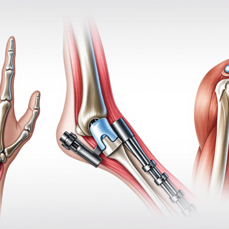
How to Choose Orthopedic Implants for Hand, Foot and Shoulder
Define the indication through anatomy before comparing implant designs. An implant is not chosen from a catalog alone. In the hand, small joints, thin soft tissue, tendons, and nearby nerves demand precise planning. Bone stock and expected motion also matter. A stable implant may suit a painful, low-demand joint, while a motion-preserving option may require stronger bone and reliable soft-tissue balance. Radiographs help, but they do not show every functional problem.
The foot carries body weight through changing positions. Alignment, joint surface damage, skin condition, and gait should guide the indication. A device that looks suitable on a standing image may still fail to address instability or abnormal loading. Foot anatomy varies greatly between patients. That makes templating useful, but not absolute.
The shoulder requires a different assessment. Humeral bone quality, glenoid version, joint congruity, and rotator cuff function can change the entire surgical plan. Instability, stiffness, fracture, and arthritis are not interchangeable indications. Clinical examination should be read alongside imaging and patient goals. I have learned that a technically elegant implant can be the wrong choice when expectations are unclear. Even careful planning has limits. Reassessment may reveal that nonoperative care, delayed surgery, or another implant concept is safer. The final decision should be documented by an experienced orthopedic team, with clear discussion of risks, rehabilitation, and realistic function.
| Anatomic Region | Relevant Anatomy | Typical Indication | Implant Category | When It Is Commonly Considered | Key Selection Factors | Important Clinical Considerations |
|---|---|---|---|---|---|---|
| Hand / Wrist | Distal radius, radial styloid, distal radioulnar joint, carpal bones, flexor and extensor tendon compartments. | Displaced or unstable distal radius fracture, especially with loss of alignment or articular incongruity. | Volarly applied fracture plate with locking or non-locking screws; selected cases may use external fixation or percutaneous pins. | When reduction cannot be maintained in a cast, the fracture is intra-articular, or early functional recovery is important. | Fracture pattern, bone quality, articular involvement, screw trajectory, tendon clearance, and restoration of radial height, inclination, and volar tilt. | Avoid excessive screw length near the extensor tendons. Median nerve symptoms, soft-tissue condition, and swelling should be assessed before surgery. |
| Hand / Metacarpals and Phalanges | Metacarpal shafts and bases, proximal and middle phalanges, collateral ligaments, extensor mechanism, and flexor tendons. | Unstable, displaced, shortened, rotated, or irreducible fractures; selected intra-articular fractures. | Mini-plates and screws, lag screws, intramedullary devices, or Kirschner wires. | When rotational alignment cannot be reliably controlled with splinting or when fracture stability is needed for early motion. | Bone diameter, fracture location, rotational deformity, implant prominence, soft-tissue coverage, and the need to protect tendon gliding. | Even small rotational errors can impair finger alignment. Prominent plates or screws may irritate tendons and sometimes require later removal. |
| Hand / Thumb Carpometacarpal Joint | Trapezium, base of the first metacarpal, saddle-shaped thumb carpometacarpal joint, and surrounding ligament complex. | Painful thumb carpometacarpal osteoarthritis with functional limitation after non-operative treatment. | Arthrodesis fixation, osteotomy fixation, or selected thumb joint replacement systems. | Arthrodesis may be considered for high-demand patients requiring stability; arthroplasty may be considered when preserving thumb motion is a priority. | Age, activity level, joint stability, bone stock, deformity, desired motion, and surgeon experience with the selected procedure. | Treatment choice should distinguish isolated carpometacarpal disease from scaphotrapeziotrapezoid arthritis or metacarpophalangeal hyperextension. |
| Hand / Finger Joints | Proximal interphalangeal, distal interphalangeal, and metacarpophalangeal joints with collateral ligaments and tendon insertions. | End-stage painful arthritis, post-traumatic joint destruction, or selected unstable joint deformity. | Silicone, pyrocarbon, or metal-polymer joint arthroplasty; arthrodesis screws or plates where fusion is preferred. | Arthroplasty is generally aimed at pain relief and motion; arthrodesis is considered when durable stability is more important than motion. | Joint level, deformity, bone stock, ligament competence, tendon balance, activity demands, and implant size. | Implant selection must account for potential instability, implant fracture or wear, silicone synovitis, loosening, and the risk of stiffness. |
| Foot / Ankle | Distal tibia, fibula, talus, ankle mortise, syndesmosis, and major ankle ligaments. | Unstable ankle fracture, syndesmotic disruption, or fracture-dislocation requiring restoration of the ankle mortise. | Anatomic plates, lag screws, intramedullary nails, syndesmotic screws, or flexible suture-button constructs. | When displacement, instability, talar shift, syndesmotic widening, or inability to maintain closed reduction is present. | Fracture classification, bone quality, soft-tissue swelling, syndesmotic stability, loading protocol, and implant prominence. | Soft-tissue swelling and skin condition can affect timing. Accurate mortise reduction is important to reduce the risk of post-traumatic arthritis. |
| Foot / Hindfoot | Calcaneus, talus, subtalar joint, talonavicular joint, and posterior facet of the calcaneus. | Displaced intra-articular calcaneal fracture, talar neck or body fracture, or hindfoot deformity requiring correction. | Low-profile plates, screws, cannulated screws, external fixation, or fusion fixation constructs. | When joint congruity, hindfoot alignment, length, or height cannot be restored and maintained non-operatively. | Soft-tissue envelope, fracture morphology from CT, weight-bearing alignment, bone quality, and risk of avascular necrosis in talar injuries. | High-energy hindfoot injuries may have substantial swelling and wound risk. Talar blood supply and postoperative weight-bearing restrictions are critical. |
| Foot / Midfoot | Tarsometatarsal joints, Lisfranc ligament, naviculocuneiform joints, cuboid, navicular, and cuneiform bones. | Unstable Lisfranc injury, displaced midfoot fracture-dislocation, or symptomatic post-traumatic midfoot arthritis. | Bridge plates, transarticular screws, low-profile plates, primary arthrodesis fixation, or percutaneous pins. | When weight-bearing alignment is lost, the Lisfranc complex is unstable, or joint surfaces are severely damaged. | Quality of reduction, ligamentous versus purely bony instability, joint preservation, bone quality, and planned weight-bearing progression. | Some transarticular fixation may require later removal. Persistent malalignment can cause chronic pain and midfoot arthritis. |
| Foot / Forefoot | First metatarsal, metatarsophalangeal joints, lesser metatarsals, proximal phalanges, sesamoids, and plantar plate structures. | Hallux valgus correction, first metatarsophalangeal arthritis, metatarsal fracture, or lesser-toe deformity. | Osteotomy plates, compression screws, intramedullary implants, arthrodesis plates and screws, or small pins. | When correction requires stable fixation, joint fusion, restoration of the first-ray position, or maintenance of metatarsal alignment. | Deformity plane, first-ray stability, bone stock, fixation compression, implant prominence, footwear demands, and bone-healing potential. | Alignment should be assessed in multiple planes. Excessive shortening or malposition can transfer load to adjacent metatarsals. |
| Shoulder / Proximal Humerus | Humeral head, surgical neck, greater and lesser tuberosities, rotator cuff insertions, and axillary nerve region. | Displaced proximal humerus fracture, fracture-dislocation, or fracture with tuberosity displacement and functional compromise. | Locking plate, intramedullary nail, screws, or hemiarthroplasty/reverse shoulder arthroplasty for selected complex fractures. | Fixation is considered when alignment is unacceptable or instability threatens shoulder mechanics; arthroplasty may be considered for unreconstructible head or tuberosity injury. | Fracture pattern, medial hinge integrity, bone quality, rotator cuff status, tuberosity repair, vascular risk, and patient age or activity level. | Monitor for screw penetration into the joint, avascular necrosis, axillary nerve dysfunction, stiffness, and tuberosity nonunion or migration. |
| Shoulder / Glenohumeral Instability | Glenoid rim, labrum, anterior capsule, humeral head, rotator interval, and inferior glenohumeral ligament complex. | Recurrent anterior instability, Bankart lesion, or glenoid bone loss requiring soft-tissue or bony stabilization. | Suture anchors, cortical button fixation, or screws and plates for coracoid transfer or other bone-block procedures. | When recurrent instability persists despite rehabilitation or when critical glenoid bone loss, engaging Hill-Sachs lesions, or high-risk activity is present. | Amount and location of bone loss, lesion tracking, soft-tissue quality, contact sports, implant position, and risk of neurovascular injury. | Three-dimensional imaging may be needed for bone-loss assessment. Improper screw placement can threaten the joint surface or nearby neurovascular structures. |
| Shoulder / Rotator Cuff and Tendon-to-Bone Repair | Greater tuberosity footprint, supraspinatus, infraspinatus, subscapularis, biceps anchor, and subacromial space. | Repairable full-thickness rotator cuff tear or selected tendon avulsion with persistent weakness and pain. | All-suture, polymer, or metal suture anchors; transosseous-equivalent or double-row fixation constructs. | When tendon quality and mobility allow restoration of the tendon footprint and symptoms remain significant after non-operative care. | Tendon retraction, fatty infiltration, tear size, bone quality, anchor pull-out strength, number and placement of anchors, and rehabilitation protocol. | Healing depends on tendon biology and rehabilitation. Avoid over-tensioning, anchor convergence, and penetration into the articular surface. |
| Shoulder / Glenohumeral Arthritis | Humeral head, glenoid surface, rotator cuff, scapular neck, and deltoid muscle. | Painful end-stage glenohumeral osteoarthritis, inflammatory arthritis, or post-traumatic joint destruction. | Anatomic total shoulder arthroplasty or reverse shoulder arthroplasty. | Anatomic replacement is generally considered with a functional rotator cuff; reverse replacement is considered when cuff function is insufficient or fracture-related reconstruction requires it. | Rotator cuff integrity, glenoid bone stock and version, deformity, deltoid function, age, activity level, and implant fixation strategy. | Infection, instability, loosening, scapular notching, periprosthetic fracture, and subscapularis or tuberosity healing must be considered during planning. |
| Cross-Regional Decision Factors | Bone, cartilage, tendon, ligament, neurovascular structures, skin, and soft-tissue envelope. | Selection of fixation, replacement, fusion, or soft-tissue fixation based on the specific pathology and functional objective. | Screws, plates, pins, nails, suture anchors, arthroplasty components, fusion constructs, or external fixation. | When the expected benefit of mechanical stability or joint reconstruction outweighs surgical and implant-related risks. | Anatomic fit, load environment, bone quality, implant rigidity, fixation purchase, imaging findings, patient goals, comorbidities, and rehabilitation requirements. | Implant choice should be individualized by a qualified orthopedic surgeon and confirmed against current clinical evidence, applicable instructions for use, and patient-specific imaging. |
Implant geometry should follow the joint’s natural motion, not merely its visible shape. In the hand, small changes in angulation can affect tendon balance and grip. A shoulder implant needs to respect rotation, elevation, and the patient’s remaining bone. Foot implants face repeated loading during walking, turning, and push-off. The design must tolerate those forces without restricting useful movement.
Bone stock changes the decision. Thin cortical bone may require broader contact or a different fixation strategy. Dense bone can provide strong support, but excessive stiffness may shift stress to nearby structures. Preoperative radiographs and advanced imaging help estimate size, alignment, and available fixation zones. During surgery, direct inspection can reveal weaker bone than imaging suggested. That moment demands judgment.
Load is not identical for every patient. A manual worker, an older adult, and a distance runner may need different mechanical priorities. Soft-tissue tension also matters. An implant can fit the bone yet disrupt a tendon’s path. Poor rotational alignment may cause pain during ordinary tasks. I have seen planning assumptions change after comparing motion under gentle stress. That is worth remembering.
There is no flawless formula. Surgeons should weigh geometry, motion, bone quality, activity level, and fixation security together. Small compromises deserve documentation and discussion. The safest choice is usually the one that preserves useful movement while respecting biological limits.
How to Choose Orthopedic Implants for Hand, Foot, and Shoulder
Material choice begins with both mechanical performance and biological safety. ASTM F136 defines requirements for wrought titanium-6Al-4V ELI used in surgical implants. Its lower density, approximately 4.4 g/cm³, can reduce implant weight. Titanium’s elastic modulus is about 110 GPa, closer to bone than stainless steel, near 190 GPa. This difference may help limit stress shielding, although implant geometry remains equally important. ASTM F136 compliance does not prove complete biological safety.
ISO 10993 requires a biological evaluation based on tissue contact and exposure duration. A temporary bone screw and a permanent shoulder component need different testing strategies. ISO 10993-1:2018 supports endpoint selection, while ISO 10993-18:2020 addresses chemical characterization. The FDA’s 2023 guidance also stresses risk-based evaluation instead of automatic testing. Review extractables, corrosion products, cleaning residues, and manufacturing particles. A polished surface can still carry invisible residues. That is an uncomfortable but practical reminder. Material certificates alone are not enough.
Tips: Ask for the exact ASTM revision, mill certificate, and ISO 10993 test matrix. Check whether testing used the finished device. Compare fatigue data, corrosion behavior, and surface treatment. Do not select titanium only because it sounds biocompatible. Anatomical load, fixation method, and patient sensitivity can change the decision. Reports from the International Organization for Standardization and FDA should support the file, not replace engineering judgment.
Material comparison under ISO 10993 biocompatibility considerations and ASTM F136 titanium alloy requirements
Titanium alloy Ti-6Al-4V ELI is the material specifically covered by ASTM F136 for wrought surgical implant applications. ISO 10993 is a series of biological evaluation standards; applicability depends on the implant’s material, contact type, contact duration and manufacturing process. Density and elastic modulus shown below are typical nominal engineering values and should be verified against the supplier’s material certification and final device testing.
Lower elastic modulus may help reduce stiffness mismatch, while final selection must also consider fatigue strength, wear, corrosion resistance, sterilization, anatomy and clinical indication.
Choosing an orthopedic implant begins with readable imaging, not a catalog. For the hand and foot, obtain standard AP, lateral, and oblique radiographs. Add weight-bearing views when alignment changes under load. For the shoulder, include true AP, axillary, and scapular-Y views. CT can clarify glenoid version, bone loss, and fracture lines. In practice, I compare both sides whenever anatomy looks unusual. Small rotational errors can alter joint mechanics and screw trajectories.
Fixation should match bone quality, contact area, and expected loading. Measure cortical thickness, defect depth, joint width, and available screw length. Do not select size from one projection. Superimposed anatomy can hide a narrow canal or underestimate a glenoid defect. The image can still mislead. A second review helps. The 2024 American Joint Replacement Registry Annual Report includes more than 3.5 million cumulative hip, knee, and shoulder procedures, showing the value of consistent imaging and longitudinal documentation. The 2024 National Joint Registry report likewise emphasizes registry-based monitoring of revision patterns and implant performance. These reports support disciplined measurement, but they cannot replace patient-specific judgment.
For hand and foot implants, preserve alignment in three planes and avoid excessive compression across fragile bone. For shoulder implants, assess humeral height, offset, inclination, and glenoid version before final sizing. Trial components should reproduce movement without over-tensioning soft tissues. Sometimes the “best” size on paper is not the safest intraoperative fit. That deserves reconsideration.
How to Choose Orthopedic Implants for Hand, Foot, and Shoulder
FDA clearance should be the starting point, not the final decision. Confirm the device’s clearance pathway, intended anatomy, materials, and surgical indications in the FDA database. Clearance confirms substantial equivalence, but it does not prove superior long-term outcomes. ISO 13485 certification shows that the manufacturer operates a controlled quality-management system. It does not guarantee every implant will perform well in every patient. That distinction matters.
Registry evidence adds real-world perspective. The Australian Orthopaedic Association National Joint Replacement Registry 2024 Annual Report analyzed more than 1.8 million procedures. Its data show that revision risk can vary by implant design, fixation method, and patient factors. Shoulder results especially require careful interpretation because reverse and anatomical replacements serve different indications. Hand and foot registries are often smaller. Their evidence may be less mature. That is a limitation worth admitting.
Tips: Ask for the latest registry report, revision rates, follow-up duration, and subgroup results. Compare outcomes for similar patients, not attractive headline numbers. Verify lot traceability, sterilization records, and surgeon training requirements. Review adverse-event signals through FDA reporting systems. A practical check is simple: can the supplier explain one unexpected failure without becoming defensive? If not, investigate further. Clinical judgment still matters. Data can be incomplete.
Start with anatomy, pain, movement, bone quality, and patient goals. Imaging helps, but it may miss tendon imbalance or functional weakness. The best implant is not always the most advanced one.
Small joints, thin soft tissue, tendons, and nearby nerves require precise planning. Minor angulation changes can affect grip and tendon movement. A stable design may suit a painful, low-demand joint.
The foot carries changing loads during standing, walking, turning, and push-off. Alignment, skin condition, joint damage, and gait should guide the decision. A standing image may not reveal instability.
Assess humeral bone quality, glenoid version, joint fit, and rotator cuff function. Fracture, stiffness, arthritis, and instability need different plans. Patient expectations matter too.
Geometry should follow useful joint motion, not only visible bone shape. The design must support movement without disrupting tendons or nearby structures. Poor rotation can cause pain during simple tasks.
Thin cortical bone may need broader contact or another fixation method. Dense bone can provide support but may transfer stress elsewhere. A manual worker and a distance runner may need different mechanical priorities.
Check regulatory clearance, intended anatomy, materials, and approved surgical indications. Review quality-system certification, revision rates, follow-up duration, and similar patient groups. Clearance does not prove superior long-term performance.
Confirm lot traceability, sterilization records, fixation requirements, and surgeon training. Ask how unexpected failures were investigated. Data can be incomplete. That is uncomfortable, but important.
Yes. Reassessment may support nonoperative care, delayed surgery, or another implant concept. I have learned that a technically elegant design can mismatch unclear expectations. Planning has limits.
Choosing the right implant begins with understanding the anatomy and clinical indication of the hand, foot, or shoulder. How to choose orthopedic implants for hand, foot, and shoulder procedures depends on the affected joint, expected motion, mechanical loading, bone quality, and available bone stock. Implant geometry should support natural joint function while promoting stability, proper alignment, and preservation of surrounding tissue. Imaging data, including radiographs or advanced scans when appropriate, can help determine implant size, positioning, and fixation strategy.
Material selection should be reviewed against biological safety and performance expectations, including ISO 10993 and ASTM F136 requirements where applicable. Clinicians should also assess cemented or non-cemented fixation, instrument compatibility, and the range of available sizes. Before use, verify regulatory clearance, manufacturing controls under ISO 13485, and relevant clinical or registry outcomes. A balanced decision combines patient anatomy, surgical goals, implant design, material safety, and reliable evidence to support durable and predictable results.

Voila Orthopedic Implants